1. Understanding Accessory Navicular Bone
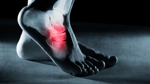
An accessory navicular bone is an extra bone or piece of cartilage located on the inner side of the foot. This bone is present at birth and can cause pain and discomfort for some individuals, especially those who are active or have flat feet.
2. Symptoms of Accessory Navicular Syndrome
Symptoms include a visible bony prominence on the inner foot, pain, and swelling. These symptoms are often aggravated by physical activity, tight shoes, or trauma to the foot, making it essential to diagnose and address the condition early.
3. Initial Diagnosis
Diagnosing accessory navicular syndrome involves a physical examination and imaging studies, such as X-rays or MRI scans. These help determine the bone’s size, shape, and its impact on surrounding tissues and structures.
4. Conservative Treatment Options
Before considering surgery, doctors usually recommend non-surgical treatments. These include rest, ice, anti-inflammatory medications, physical therapy, and custom orthotics to relieve pressure on the affected area.
5. When Surgery Becomes Necessary
Surgery is considered when conservative treatments fail to alleviate pain and improve function. Persistent pain, limited mobility, and significant impact on daily activities are key indicators that surgery might be required.
6. Pre-Surgery Preparations
Preparation for surgery involves a thorough medical evaluation, including blood tests and imaging. Patients should discuss their medical history, current medications, and any allergies with their surgeon to ensure a safe procedure.
7. The Surgery Procedure
Accessory navicular bone surgery typically involves removing the extra bone and reshaping the surrounding area to relieve pain and improve foot function. The surgery is performed under general or local anesthesia, depending on the patient’s condition.
8. Immediate Post-Surgery Care
After surgery, patients are usually required to keep the foot elevated and apply ice to reduce swelling. Pain management is crucial, and medications are prescribed to control post-operative discomfort.
9. The Importance of Rest
Rest is vital in the initial recovery phase. Patients should avoid putting weight on the affected foot and use crutches or a walker as recommended by their surgeon to facilitate healing.
10. Physical Therapy Begins
Physical therapy often starts a few weeks after surgery. Therapists work with patients to restore range of motion, strengthen the foot, and gradually return to normal activities without risking re-injury.
11. Monitoring Progress
Regular follow-up appointments with the surgeon are essential to monitor healing and address any concerns. X-rays or other imaging tests may be performed to ensure the bone and surrounding structures are healing correctly.
12. Potential Complications
As with any surgery, there are potential complications, including infection, blood clots, and nerve damage. However, these are rare and can usually be managed effectively with prompt medical attention.
13. Pain Management Strategies
Effective pain management includes both medications and non-pharmacological methods such as ice therapy, elevation, and compression. It’s crucial to follow the surgeon’s advice for optimal pain control.
14. Gradual Return to Activities
Returning to normal activities should be gradual. Patients should follow their physical therapist’s guidance and avoid high-impact activities until the foot is fully healed to prevent setbacks.
15. Long-Term Foot Care
Long-term care involves wearing supportive footwear, using custom orthotics if recommended, and maintaining a healthy weight to reduce stress on the feet. Regular foot exercises can also help maintain strength and flexibility.
16. Patient Experiences
Hearing from other patients who have undergone the surgery can be reassuring. Many report significant pain relief and improved mobility, highlighting the surgery’s potential benefits.
17. Psychological Impact
The psychological impact of living with chronic foot pain can be significant. Post-surgery, many patients experience improved mood and quality of life due to reduced pain and increased mobility.
18. Children’s Recovery
Children generally recover faster than adults due to their resilient tissues and bones. However, ensuring they follow post-surgery care instructions is crucial for a smooth recovery.
19. The Role of Nutrition
Proper nutrition supports healing. A balanced diet rich in vitamins, minerals, and protein can aid in tissue repair and overall recovery after surgery.
20. Sports and Physical Activities
For athletes, returning to sports requires a careful, phased approach. Physical therapists and doctors work together to create a plan that ensures a safe return to physical activity.
21. Importance of Follow-Up Care
Continued follow-up care ensures that any issues are promptly addressed. This includes monitoring for signs of re-injury or complications and making necessary adjustments to treatment plans.
22. Cost of Surgery
The cost of accessory navicular bone surgery can vary based on factors such as location, surgeon’s fees, and insurance coverage. Patients should discuss these aspects with their healthcare provider beforehand.
23. Insurance Considerations
Insurance coverage for the surgery varies. It’s important to check with your insurance company to understand what is covered and what out-of-pocket expenses you might incur.
24. Post-Surgery Support Network
Having a support network is beneficial. Family and friends can assist with daily tasks during the recovery period, helping patients focus on healing and rehabilitation.
25. Looking Forward
Post-surgery, many patients look forward to returning to their favorite activities without pain. With proper care and rehabilitation, the outcome of accessory navicular bone surgery can be very positive, leading to a more active and pain-free life.